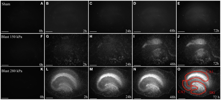
Figure 3.
Cell death in OHCs after blast exposure. Following the 8 DIV recovery period from dissection, OHCs were exposed to a 150 kPa (low) or 280 kPa (high) blast overpressure or were sham-injured. (A–E) Representative micrographs of sham OHCs over a time course of 72 h, demonstrating low levels of dead PI-stained cells (white) throughout the experiment. In OHCs exposed to a low (F–J) or high blast (K–O) overpressure, dead cells (white) were observed as early as 2 h following injury and the damage intensified at later time points. The CA1, CA3, and the DG hippocampal regions [outlined in red in (O)] appear particularly vulnerable to the blast in both high and low groups. Scale bars 500 μm.

