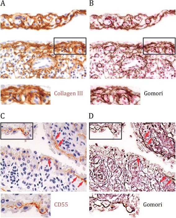
Figure 2.

Expression pattern of CD55 and collagen III coincides with collagenous structures in rheumatoid arthritis (RA) synovial tissue. Sections of RA synovial tissue first were stained with (A) anti-collagen antibody or (C) anti-CD55 antibody and then processed to (B/D) Gomori silver impregnation. Red arrowheads indicate localization of CD55 to collagenous fibers. Shown are representative stainings derived by light microscopy; magnification 20 x.
