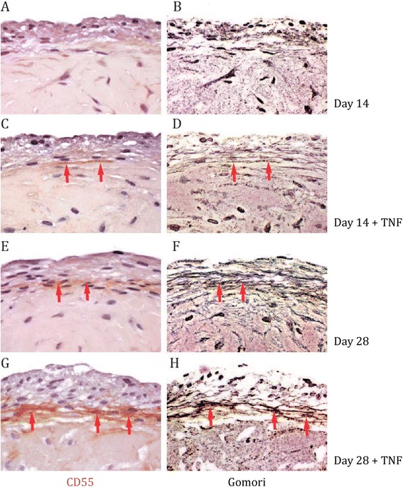
Figure 3.

Expression pattern of CD55 coincides with collagenous structures in three-dimensional fibroblast-like synoviocytes (FLS) micromasses. (A, B, E, F) Sections of three-dimensional FLS micromasses were stained with anti-CD55 antibody at day 14 (A) and day 28 (E), and then processed to Gomori silver impregnation (B and F, respectively). (C, D, G, H) Sections of three-dimensional FLS micromasses, which were cultured with 10 ng/ml tumor necrosis factor (TNF), were stained with anti-CD55 antibody at day 14 (C) and day 28 (G), and then processed to Gomori silver impregnation (D and H, respectively). Red arrowheads indicate a similar distribution of CD55 and collagenous fibers. Shown are representative stainings derived by light microscopy; magnification 40 x.
