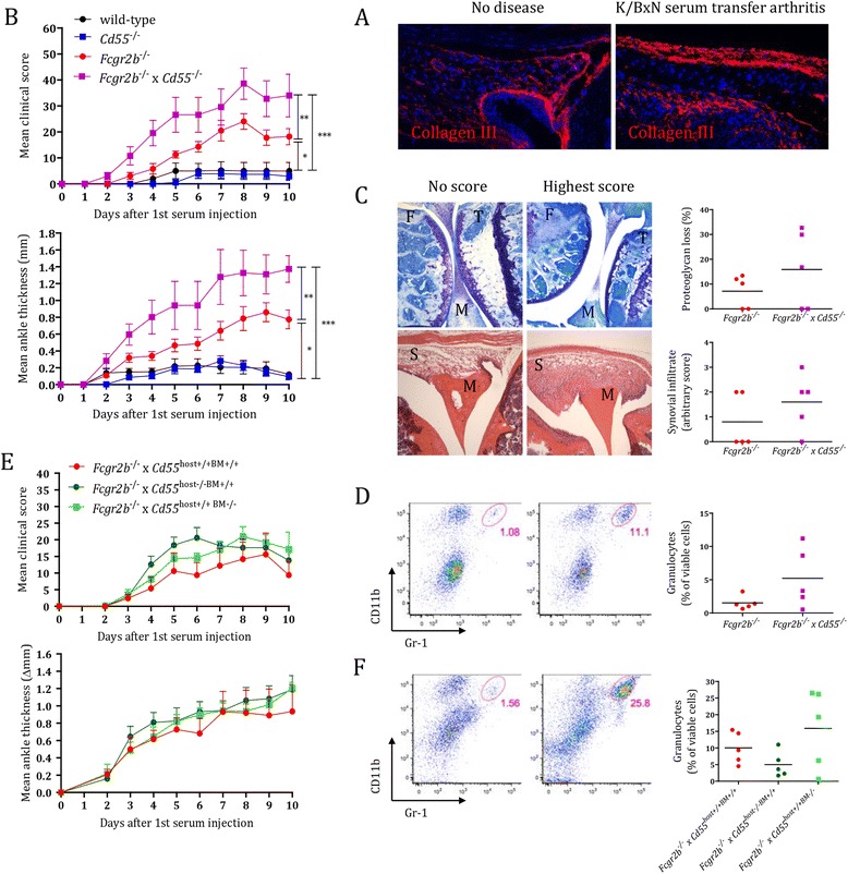
Figure 5.

CD55 either on immune or stromal cells protects from K/BxN serum transfer-induced arthritis in the absence of FcγRIIB (CD32). Mice were injected at day 0 with 12.5 μl/g K/BxN serum and followed for disease development. At day 10, animals were sacrificed for further analysis. (A) Collagen III staining in representative knee joint sections of non-diseased and arthritic Fcgr2b −/− mice. Magnification 20 x. (B) Development of arthritis evaluated by measuring clinical scores and ankle thickness. Depicted are mean ± standard error of the mean (SEM) of five animals per group. ***, P <0.001; **, P <0.01; *, P <0.05 (C) Histological evaluation of knee joints for proteoglycan loss (toluidine blue; above) and cell infiltration (hematoxylin and eosin; below). Photographs are representative of mice with no or highest disease activity in the experiment, respectively. Joint structures are labeled as F, femur; M, meniscus; S, synovium; T, tibia. Magnification 5 x. (D) Flow cytometry plots of isolated synovial tissue cells stained for CD11b+Gr1+ granulocytes, representative of mice with no or highest disease activity in the experiment, respectively. (E) Development of arthritis evaluated by measuring clinical scores and ankle thickness in bone marrow chimeric mice that lacked CD55 on stromal cells (Fcgr2b −/− x Cd55 host−/−BM+/+, depicted as a dark green line), on immune cells (Fcgr2b −/− x Cd55 host+/+BM−/−, depicted as a light green line), or on none of the two compartments (Fcgr2b −/− x Cd55 host+/+BM+/+, depicted as a red line). Depicted are mean ± SEM values of five animals per group. (F) Flow cytometry plots of isolated synovial tissue cells stained for CD11b+Gr1+ granulocytes, representative of mice with no or highest disease activity in the experiment, respectively. Quantifications provided to the right in (C), (D), and (F) show individual mice (dots) with a horizontal line indicating the mean per group (n = 5).
