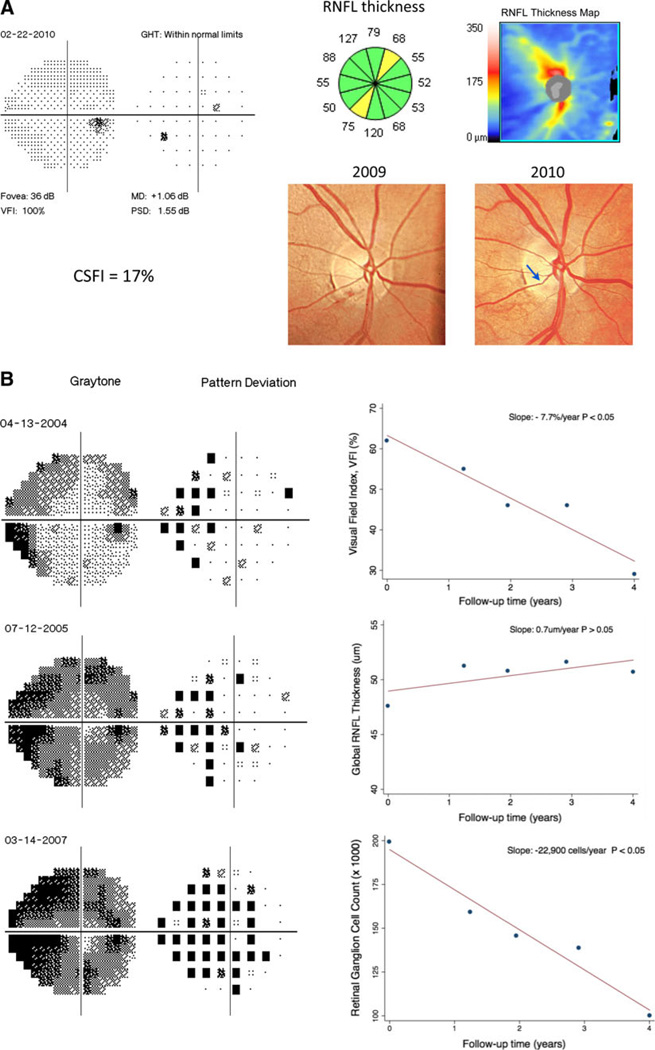
Fig. 1.
Examples of detection of glaucomatous damage and estimation of rates of disease progression using a combined structure and function approach. a Example of an eye with pre-perimetric glaucomatous damage. The eye had evidence of progressive neuroretinal rim thinning over time (arrow) with corresponding RNFL loss on SD-OCT. The visual field exam still had all parameters within statistically normal limits. The CSFI was 17 %, indicating a loss of 17 % of RGC compared to the age-expected normal number. b Example of an eye showing severe glaucomatous damage and progressive visual field loss over time. The parameter visual field index showed a deterioration of −7.7 %/year (P < 0.05). Despite the clear visual field progression, longitudinal evaluation with SD-OCT did not show change in RNFL parameters. The use of a combined approach to evaluate the rate of RGC loss, however, estimated a decline of −22,900 cells/year, which was statistically significant (P < 0.05). RNFL retinal nerve fiber layer, CSFI combined structure and function index, VFI visual field index, MD mean deviation, PSD pattern standard deviation, GHT glaucoma hemifield test

