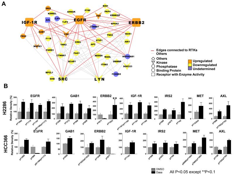
Fig. 3. Increased tyrosine phosphorylations in receptor tyrosine kinase (RTK) after dasatinib exposure.
A: Protein-protein interaction (PPI) network composed of dasatinib-regulated pY proteins identified from both H2286 and HCC366 cells. Each protein was annotated based on its functional classification, with PPI then mapped utilizing BisoGenet Cytoscape plug-in. Increased tyrosine phosphorylation of EGFR, IGF-1R, and ERBB2 and decreased tyrosine phosphorylation of SRC and LYN are presented in bigger nodes. Edges connected to EGFR, IGF-1R and ERBB2 are shown in red, with others in gray. “Undetermined” signifies pY peptides showing conflicting direction (increased versus decreased) in the two cell lines. B: Extracted ion chromatogram (EIC) for pY peptides corresponding to RTK. Two-sample t-test was performed to address significant difference in EIC between control and dasatinib treatment.

