
Fig 1. S. Typhimurium fails to elicit MC activation and subsequent neutrophil recruitment and bacterial clearance in vivo.
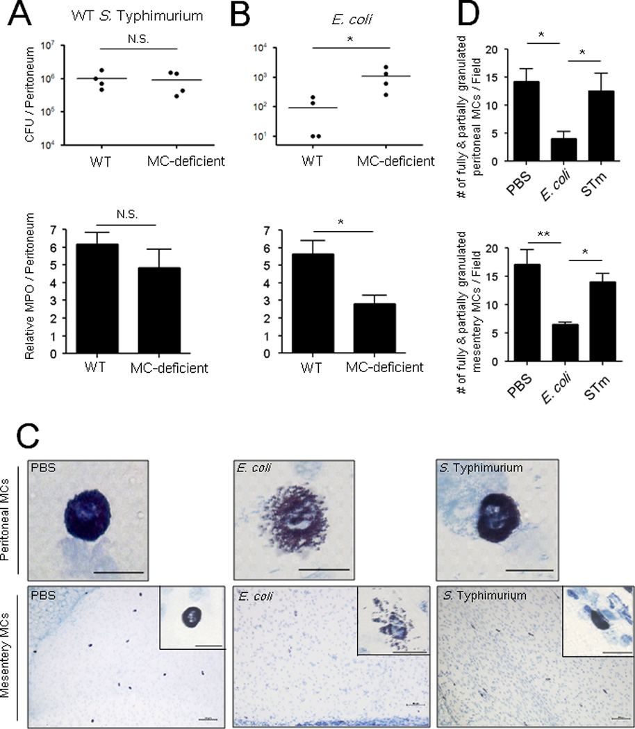
(A, B, top) Residual bacterial counts (CFUs) in the peritoneal cavity of wild-type (WT) or MC-deficient mice 24 h following intraperitoneal (i.p.) infection with 5×10⁵ CFU S. Typhimurium or 1×10⁷ CFU E. coli. (A, B, bottom) Myeloperoxidase assay of peritoneal lavage of WT or MC-deficient mice 5 h post-infection with 1×10⁷ CFU S. Typhimurium or E. coli J96 (C) Morphologic appearance of MCs in the peritoneum (top) and mesentery (bottom) of WT mice 3 h after i.p infection of 1×10⁸ CFU S. Typhimurium or E. coli J96. Insets represent 60X magnification of MCs in the mesentery. Images represent 3 independent experiments. Top panels and inset scale: 20 µm. Bottom panels scale: 100 µm. n=4. (D) Granulated MC numbers in peritoneal lavages (top) or mesentery whole mounts (bottom) were quantified by counting the number of partially and fully granulated MCs/field (n=3–5; 5 random chosen fields). Wholly degranulated MCs could not be detected. Mean ± SEM, *p<0.05, **P<0.01, N.S., not significant.