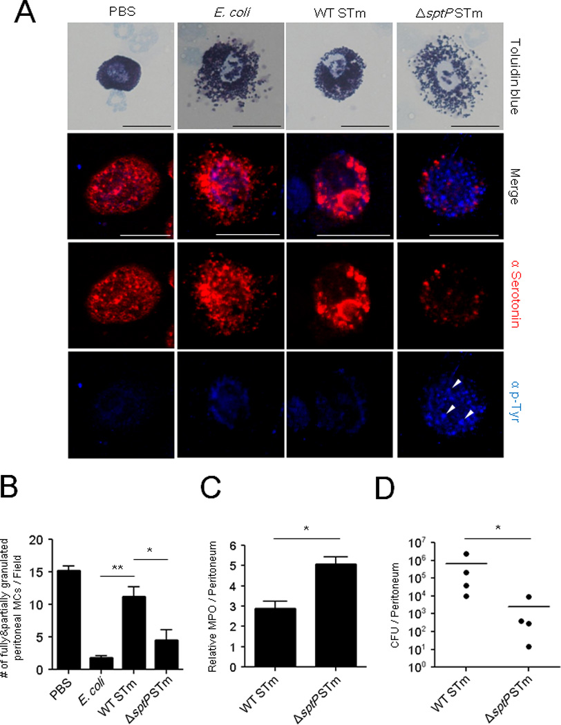
Fig 5. Infection of mice with ΔsptP activates MCs and enhances neutrophil recruitment and bacterial clearance compared to WT.

(A) WT, ΔsptP STm, or E. coli J96. 1×10⁸ CFU WT, ΔsptP STm, or E. coli J96 were injected i.p. Peritoneal lavage was harvested 4 h later, cytospun, and stained with toluidine blue for bright field microscopy (top) or with α-serotonin (red) for granules, and α-phosphotyrosine (blue) for immunofluorescence microscopy. (B) Granulated MC numbers in peritoneal lavages of (A) were quantified by counting partially and fully granulated MCs/field (n=3–6; 5 random chosen fields). Wholly degranulated MCs could not be detected. (C) Assays for neutrophil recruitment (MPO) and (D) bacterial clearance (CFUs) were performed on lavages collected 4 h and 24 h, respectively, after i.p injection of 1×10⁶ CFU WT or ΔsptP STm. n=4 mice, mean ± SEM, *p<0.05, **p<0.01. Scale bar: 20 µm. See also Figure S3.
