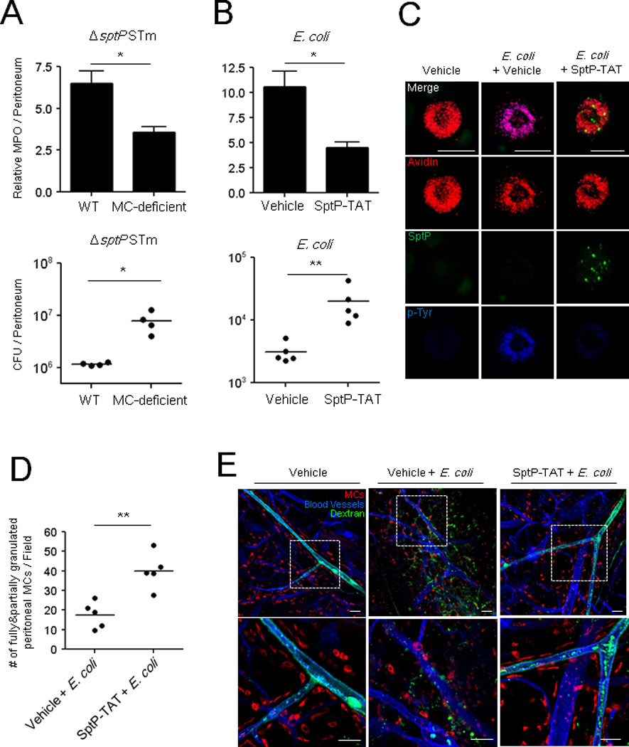
Fig 6. ΔsptP S. Typhimurium evokes MC-dependent neutrophil recruitment, bacterial clearance and vascular leakage.

Neutrophil influx (top) and bacterial counts (bottom) in WT and MC-deficient mice (A) following i.p. injection of 5×10⁵ CFU ΔsptP STm, (B) SptP-TAT (100 µg/mouse) or vehicle followed 30 min later with 1×10⁷ CFU E. coli. MPO assays were performed 5 h post-infection and CFUs 24 h post-infection. (C) Morphology of MCs in the peritoneal cavities of control and SptP-TAT treated mice 3 h following E. coli infection. MC granules: avidin (red), SptP-TAT: anti-His6 (green) and sites of tyrosine phosphorylation: anti-p-Tyr antibodies (blue). Scale bars: 20 µm. (D) Granulated MC numbers in peritoneal lavages of (C) were quantified by counting the number of partially and fully granulated MCs/field (n=5; 5 random chosen fields). Totally degranulated MCs could not be detected. (E) Mouse ears were injected intradermally with vehicle or SptP-TAT (20 µg) and injected 1 h later with 1×10⁶ CFU E. coli J96 at the same site and FITC-dextran (green) i.v.1 h post-infection, the ears were dissected, stained with avidin (red) and anti-CD31(blue), and whole mounted for immunofluorescence microscopy. Mean ± SEM, *p<0.05, **p<0.01. Scale bars: 50 µm. See also Figure S4, S5, and S6.
