
Figure 1. Self-assembly of PMI CAs into supramolecular ribbons.
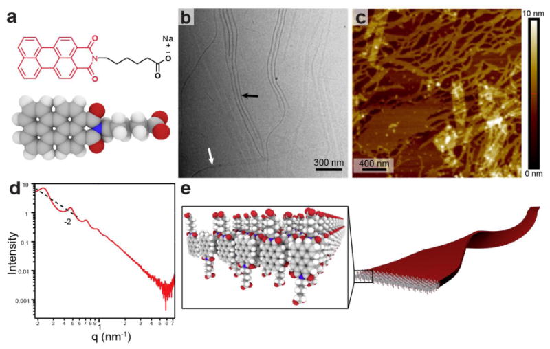
a, Molecular structure and space filling model of PMI-based CA, b, Cryo-TEM micrograph of CA solution (11.5 mM) reveals 40 nm wide supramolecular ribbons. Differences in contrast arise from imaging a ribbon either face-on (white arrow) or edge-on (black arrow). c,AFM image of a dilute CA solution (115 μM) dried on mica shows flat structures with a measured height of 2.3±0.4 nm. d,SAXS profile for aqueous CA solution (11.5 mM) exhibits a -2 slope in the low q region as well as scattering peaks corresponding to a 27.3 nm lamellar spacing between nanostructures. e, Schematic representation of the anti-parallel packing of CA molecules into a highly interdigitated bilayer to form supramolecular ribbons.