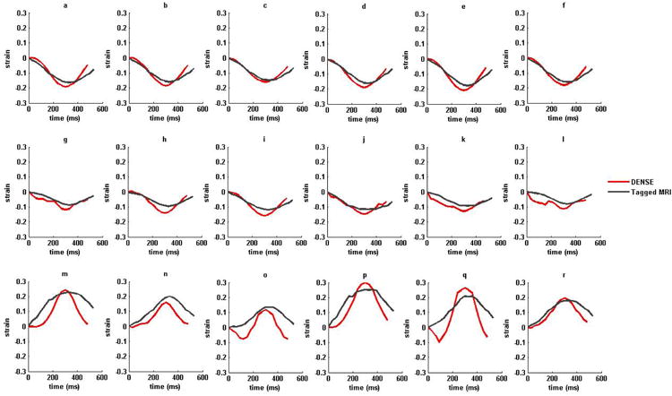
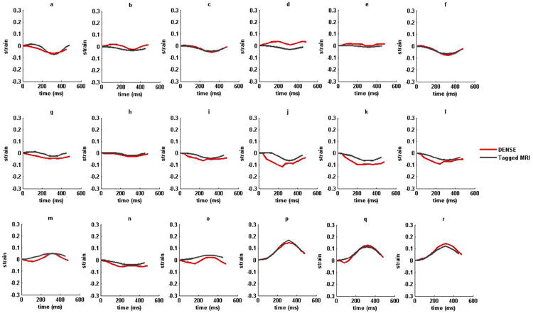
Figure 7.


(i) Strain versus time plots for sixteen AHA recommended LV segments from a normal subject in (a)–(f) circumferential (g)-(l) longitudinal and (m)-(r) radial strains. (ii) Strain versus time plots for sixteen AHA recommended LV segments from a non-ischemic, non-valvular dilated cardiomyopathy (HF) patient in (a)–(f) circumferential (g)-(l) longitudinal and (m)-(r) radial strains. Strains estimated with DENSE are shown in red and tagged MRI in dark-gray. The six LV regions by column from left to right are anterior, anteroseptal, posteroseptal, posterior, posterolateral and anterolateral in both DENSE and TMRI.
