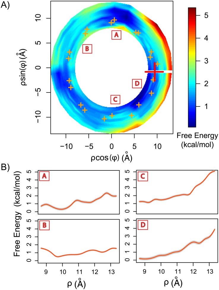
Figure 4. Binding free-energy landscape of methyl-guanidinium in the minor groove.
A) PMF was computed for one complete turn (2π) along the minor groove in the helical coordinates system (see S1 Fig.). Notice that in this coordinate system there is no angular periodicity. The PMF wasprojected to the 2D plane, such that the z-axis is perpendicular to the page. Orange crosses correspond to the average position of the DNA’s backbone phosphates of the studied turn (i.e. 10.5 base pairs). The red cross correspond to the methyl-guanidinium initial center of mass position. B) 1D-PMFs of removing the methyl guanidinium from the minor groove at 4 different angular positions (red letters in boxes) as shown in A. Grey shadow correspond to one standard deviation.

