
Fig. 5.
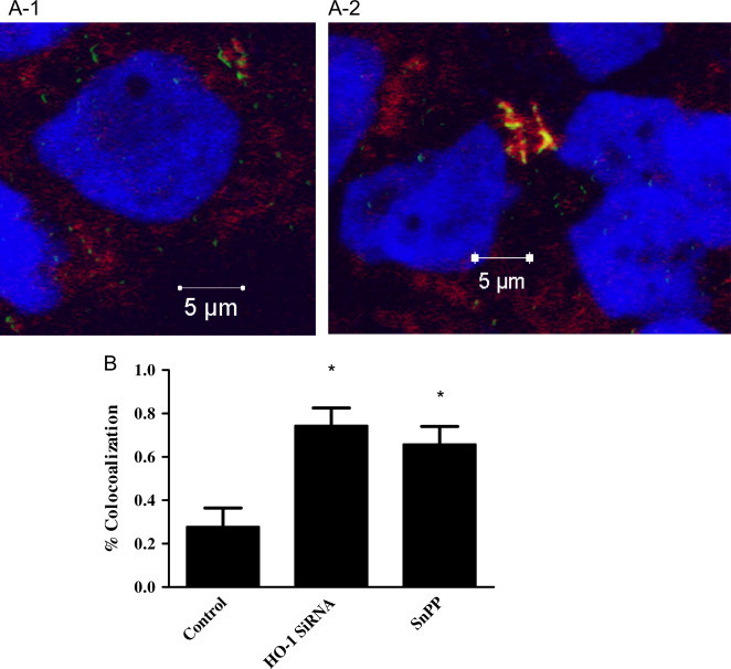
HO-1 inhibition increases M.abs–lysosomal fusion in macrophages. TPA-differentiated THP-1 cells were cultured in the presence of SnPP or HO-1 siRNA for 24 h. Macrophages were infected with Syto-62 labeled M.abs for 1 h, and incubated for 4 h. Lysotracker was added 2 h before preparation for confocal microscopy. (A-1) control cells infected with M.abs (shown in green), lysotracker is shown in red, and nucleus in blue. (A-2) M.abs infection in the presence of HO-1 siRNA. M.abs–lysosomal co-localization is shown in yellow shows (n=5). (B) Percent co-localization in SnPP and HO-1 siRNA treated macrophages as compared to control. HO-1 inhibition resulted in a significant increase in the percentage of M.abs showing co-localization with lysosomes (n=6, p<0.05).