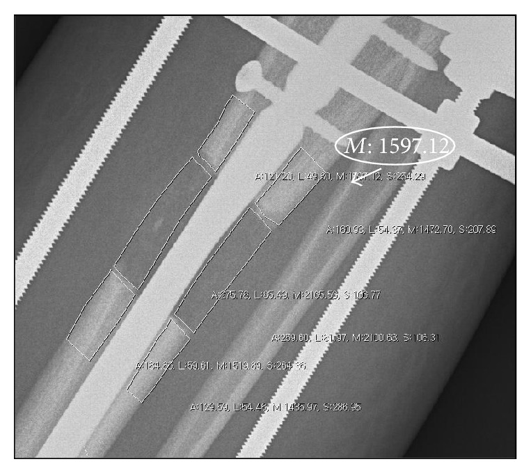
Figure 2.

The radiograph shows how to measure the pixel values on a digital radiograph. The different cortical segments of callus and the proximal and distal segments were measured with use of the free region of interest methods of StarPACS PiView Star 5.0.6.1 software (Infinitt Co., Ltd., Seoul, Korea). White circled “M” value was the pixel value of the region of interest drawn.
