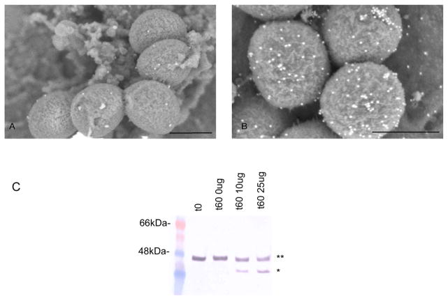
Figure 2. AniA is expressed on the cell surface in N. gonorrhoeae 1291.
Examination of N. gonorrhoeae 1291 cells grown anaerobically immunolabelled with (A) pre-immune rabbit serum or (B) anti-AniA polyclonal rabbit serum using SEM. Scale bars indicate 500nm. (C). Western blot analysis of whole, intact anaerobicaly grown N. gonorrhoeae 1291 cells treated with increasing concentrations of trypsin using anti-AniA polyclonal rabbit serum. ** = Full-length AniA, * = Digested AniA. No significant differences between the CFUs/ml at t0 and at 60mins for each of the samples were detected (Supplementary Table 3).

