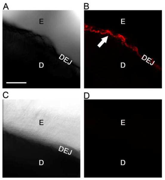
Figure 6. Confocal microscopy reveals MMP-20 is enriched in the dentin enamel junction within mature human teeth.
A, Ten micron thick stacked bright field image (10X) of a demineralized cropped crown section immunostained for MMP-20, showing enamel (E), dentin (D) and dentin enamel junction (DEJ). B, Fluorescent field image (10X) of same section shown in A showing that MMP-20 (pseudo colored in red) is strongly immunostained at the DEJ (white arrow). Weaker reactivity is evident in the adjoining enamel matrix and adjacent dentin tubules. C, Ten micron thick stacked bright field negative control image (10X) of a demineralized crown section immunostained similarly but with purified non-immune rabbit IgG. D, Fluorescent field image (10X) of the crown section shown in C showing no staining. Scale bar=100 microns (all images same magnification).

