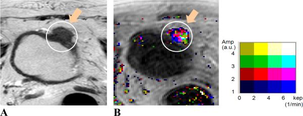
Figure 2.

MR images of a 64 year-old male patient. Image A, axial T2W image; Image B, Amp+kep map. The patient was treated with chemotherapy. Tumor location (indicated by orange arrows and enclosed in white contours) was at the anterior and dome aspect of the bladder wall. Tumor stage: T3b; size: 38 mm. The malignant tumor was visualized on both T2W image and pharmacokinetic map. The malignancy was identified with continuous color pixels on the color DCE map.
