
Figure 4.
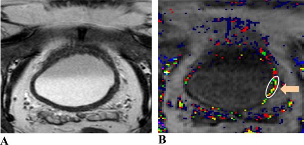
MR images of a 68 year-old male patient. Image A, axial T2W image; image B, Amp+kep map. The patient was treated with chemotherapy. Tumor location (indicated by an orange arrow and enclosed in a white contour) is located at the left and posterior aspect of the bladder wall. Tumor stage: T2; size: 9 mm. The malignant tumor with a smooth margin was not visualized on the T2W image. The malignancy was identified with continuous color pixels on the DCE-MRI map. This malignancy was both sub-centimeter and within the bladder wall thickening.