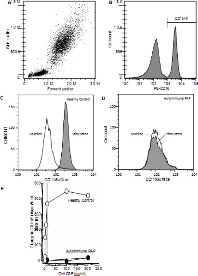
Fig. 1.
Evaluation of CD11bSurface on the neutrophil in the whole blood: an assay to measure the GM-CSF-stimulated increase in cell-surface CD11b on neutrophils in whole blood. A. Representative leukocyte cytogram. Whole blood was processed and evaluated by flow cytometry as described in the Methods. B. Identification of neutrophils by gating on CD16. Immunostained leukocytes were gated for phycoerythrin (PE)-fluorescence to identify neutrophils as a distinct CD16Hi population. C-D. Quantification of neutrophil cell-surface CD11b (CD11bSurface) for a healthy control (C) and a patient with autoimmune PAP (D). Representative histogram of the fluorescence intensity in neutrophils from healthy control. open area – no GM-CSF stimulation; filled area – after GM-CSF stimulation. E. Percent change in CD11bSurface for healthy individual (HC) and patient with autoimmune pulmonary alveolar proteinosis (PAP).

