Figure 1.

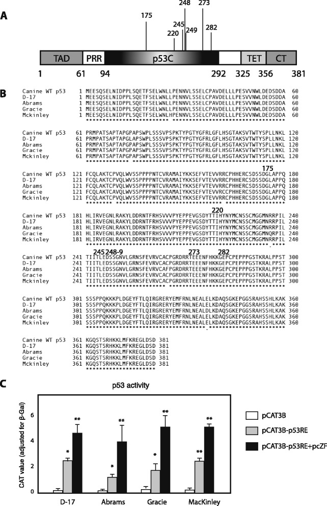
p53 in dog osteosarcoma cell lines. (A) Schematic structure of full-length p53. TAD: N-terminal transactivation domain; PRR: proline-rich region; p53C: central DNA-binding domain; TET: tetramerization domain; CT: extreme carboxyl terminus. p53C is the domain where most cancer-associated p53 mutations are located. The numbers below the diagram indicate amino acid residues delineating the domains and numbers above the diagram represent the residues with highest frequency of oncogenic missense mutations [20]. (B) Derived amino acid sequence alignment of p53s from 4 dog OS cell lines and dog wild-type p53. The residues that have high mutant frequency were marked above the diagram. Accession numbers: KP279761, KP279762, KP279763, KP279764 (C) p53 proteins of dog OS cell lines have transcriptional activity, and Zhangfei enhances p53-dependent transactivation. D–17, Abrams, McKinley, and Gracie cells were transfected with 0.5 μg of pCAT3B or pCAT3B-p53RE, in the presence or absence of 1 μg of pcZF. 24 h after transfection, the CAT activity was determined. Values represented the relative CAT activity (adjusted by β-galactosidase) of different treatments. Standard deviations from means of three individual experiments are shown. Significance of differences of the means (*P < 0.05, **P < 0.01) were determined using ANOVA.
