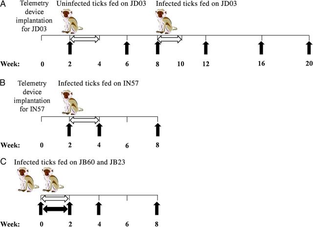
Figure 1.
Experimental design for infecting rhesus macaques with Borrelia turicatae. A and B, JD03 (A) and IN57 (B) were implanted with radiotelemetry devices 2 weeks before ticks fed on the animals. C, After the feeding, body temperatures of the 2 animals were recorded manually (black horizontal arrow) in 24-hour intervals. Horizontal white arrows indicate the time line for collecting blood specimens by ear or tail prick, and black vertical arrows indicate time points for serum specimen collection and performing complete blood count and blood chemistry analyses.

