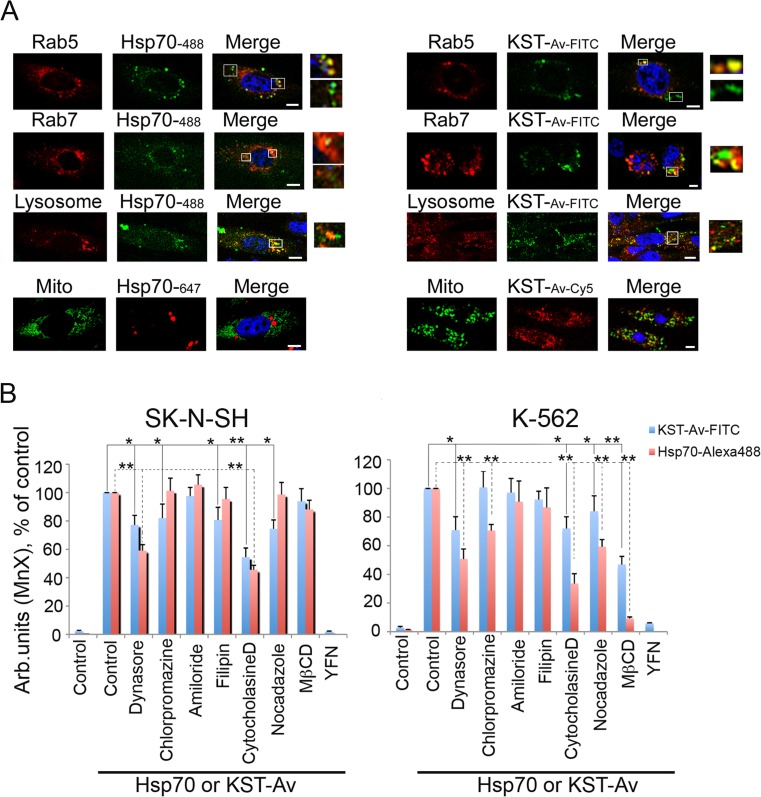
Fig. 5.
KST–Av complex penetrates inside living cells using different mechanisms. a SK-N-SH cells were transfected with plasmids bearing either rab5-RFP (early endosome marker) or rab7-RFP (late endosome marker) or stained with Lysosomal Staining Kit, revealing next step of endocytosis; when endosomes or lysosomes became visible, KST–Av–FITC or Hsp70-Alexa488 were introduced into the culture medium. Additionally, SK-N-SH cells transfected with mito-PAGFP plasmid (GFP-based mitochondrial marker) incubated with both polypeptides were studied with the aid of confocal microscopy. Scale bar 1 mm. b SK-N-SH cells (left) and K562 cell (right) were incubated with KST–Av–FITC or Hsp70-Alexa488 in the presence of intracellular transport inhibitors and investigated with the help of flow cytometry. Data from three independent experiments are summarized. Differences between control and treatment groups were considered to be statistically significant when p < 0.05 (*) or p < 0.01 (**) according to Student’s t test

