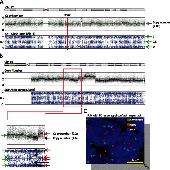
Figure 3.

Detection of polyploidy. (A) SNP and copy number data across chromosome 17 from tumor sample 5. The top panel displays the copy number probe intensity calls and the calculated copy number segments (in color). The calculated segment (green line) has an intensity value of just over 2. The lower panel displays the calculated SNP allele ratios and shows that the entire chromosome 17 is in allelic balance. The vertical red line indicates the position of HER2. (B) SNP and copy number data across chromosome 14 from tumor sample 5. The enlargement of the red box shows that a segment (green line) is predicted with an intensity value of just under 2. However, a weak allelic imbalance (green arrows) suggests that the intensity value of just under 2 does not correspond to 2 DNA copies. Moreover, a deletion (~1.6) and an amplification (~2.2) only result in a modest copy number intensity change. Taken together, the data in (A) and (B) suggest that a segment with an intensity value of just over 2 and allelic balance must correspond to at least 4 copies of DNA. (C) Representative image of a 3D-rendered model of a confocal image stack of a section from tumor sample 5 hybridized with HER2 (red) and CEP17 (green) probes. The image extends 6 μm down into the z-axis, corresponding to ~60–70% of the nucleus diameter.
