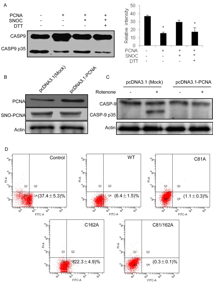
Fig 5. Regulation of caspase-9 activity by the S-nitrosylation status of PCNA.
(A) Recombinant wild-type PCNA was immobilized onto nickel-agarose beads followed by incubation with and without SNOC. After the incubated beads were added to the HeLa cytosolic extracts, the activity of caspase-9 cleavage was measured by Western blot using an anti-caspase-9 antibody. As a control, DTT was used to reduce the S-nitrosylation of PCNA induced by SNOC. The relative intensities of cleaved caspase-9 are shown in the bar chart (n = 3, *P<0.05 versus blank group). (B) Western blot analysis to PCNA overexpression and S-nitrosylation status in SH-SY5Y cells that were transfected with pcDNA3.1-PCNA. (C) Caspase-9 cleavage assay in SH-SY5Y cells with or without PCNA overexpression. The cells were treated with 500 nM rotenone for 16h followed by the cleavage activity assay based upon Western blot using antibody against caspase-9. (D) Annexin V-FITC/PI dual staining assay for rotenone-induced apoptosis in the SH-SY5Y cells which were WT, C81A, C162A, and C81/162A PCNA overexpressed, respectively. The apoptotic rates are shown as the average ratio ± SEM (n = 3).

