Figure 1.

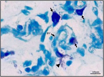
Intact (arrow) and degranulated (arrow head) MCs around a small blood vessel in the dermis. BV-blood vessel, toluidine blue stain, 400× magnification, Bar = 10 μm.

Intact (arrow) and degranulated (arrow head) MCs around a small blood vessel in the dermis. BV-blood vessel, toluidine blue stain, 400× magnification, Bar = 10 μm.