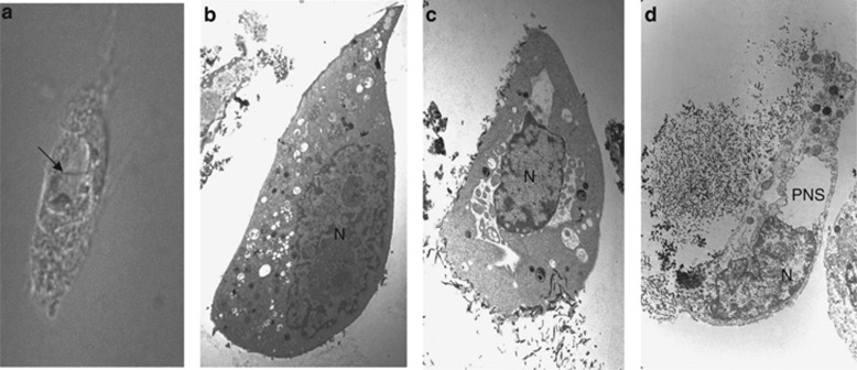
Figure 2.
Morphological features of autosis. (a) Representative light microscopic image of an autotic HeLa cell during starvation. Arrow shows area of focal nuclear concavity with adjacent focal ballooning of perinuclear space. (b–d) Representative electron microscopic images of different stages of autotic cell death; including (b) a cell in an early stage of autosis (referred to as phase 1a) with nuclear membrane convolution, mild chromatin condensation, numerous autophagosomes, and autolyososomes, dilated and fragmented ER, and electron-dense mitochondria; (c) a cell in a mid-stage of autosis (referred to as phase 1b) with separation of the inner and outer nuclear membrane, the presence of membrane-bound densities in the perinuclear space; and (d) a cell in the final stage of autosis (referred to as phase 2) with focal nuclear concavity, focal ballooning of the perinuclear space (which is empty) and disappearance of cellular organelles such as ER, autophagosomes, and autolysosomes. N, nucleus; PNS, perinuclear space

