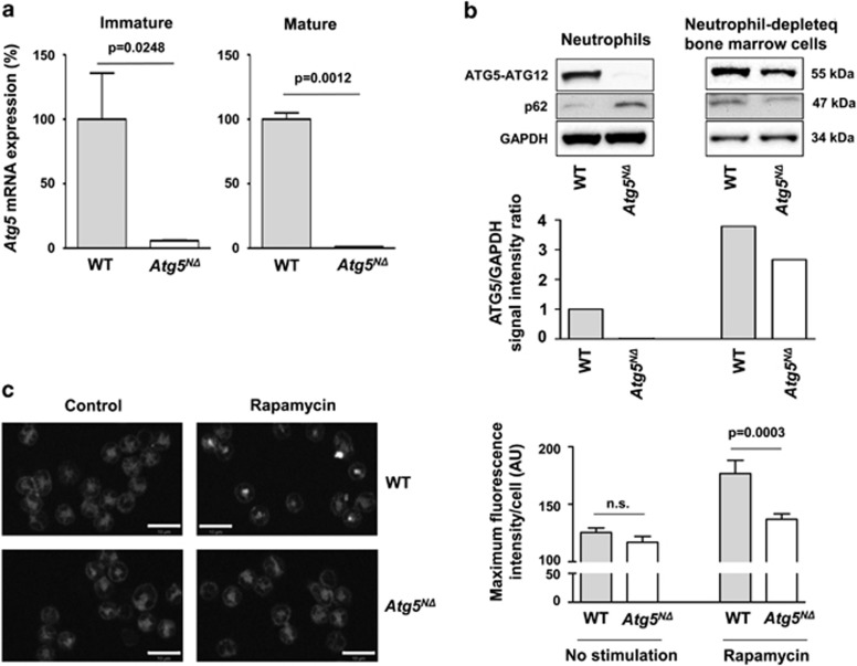
Figure 1.
Lack of Atg5 mRNA and ATG5 protein expression as well as a functional defect in autophagy in neutrophils of Atg5NΔ mice. (a) Quantitative real-time PCR. Freshly purified immature and mature bone marrow-derived neutrophil samples from WT and Atg5NΔ mice were analyzed for Atg5 expression. S18 was used as a reference gene to normalize the expression of Atg5. Values are means±S.E.M. of two independent experiments. (b) Immunoblotting. Freshly purified mature bone marrow-derived neutrophils of WT and Atg5NΔ mice were analyzed for ATG5 protein expression. The neutrophil-depleted bone marrow samples were lysed and immunoblotted next to the neutrophil lysates to demonstrate the cell specificity of the ATG5 knockout. If present, ATG5 protein is seen as a conjugate with ATG12. The accumulation of p62 confirms the attenuated autophagic activity in ATG5-deficient neutrophils. The results are representative of three independent experiments. The quantification of the ATG5 signal was performed using the Odyssey Fc Imaging System (LI-COR Biosciences GmbH, Bad Homburg, Germany) and the image Studio software. (c) Confocal microscopy. Freshly purified mature bone marrow-derived neutrophils of WT and Atg5NΔ mice were cultured in the presence and absence of rapamycin for 1 h and stained with AUTOdot autophagy visualization dye. The dye is specific for autophagic vacuoles that can be detected in rapamycin-treated WT neutrophils as bright white dots. In contrast, rapamycin had no detectable effect on neutrophils derived from Atg5NΔ mice. Bars, 10 μm. (Right) Statistical analysis of three independent confocal microscopy experiments. Values are means±S.E.M.

