Figure 3.
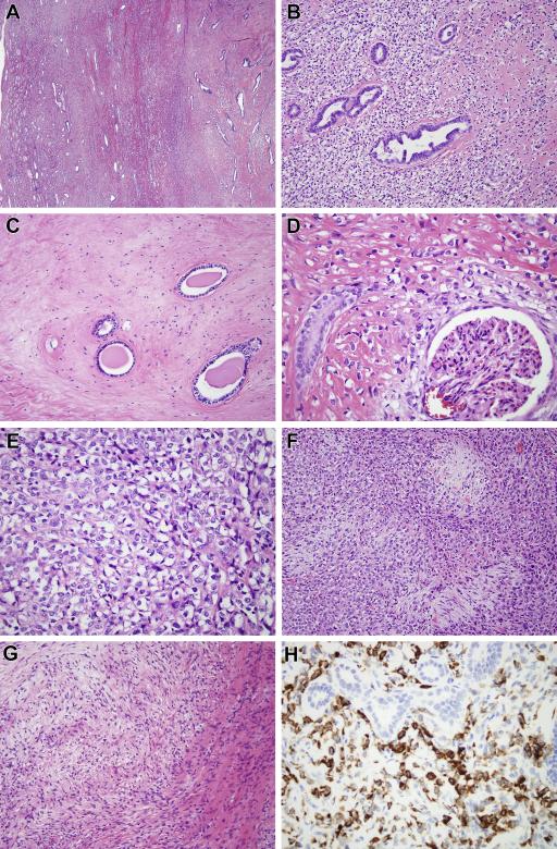
(Case 1) A. Low power view shows the spindle cell neoplasm centered within the kidney and entrapping renal tubules at its periphery. B. The neoplasm consists of epithelioid cells with clear cytoplasm, associated with stromal hyalinization. The neoplasm entraps native renal tubules, which show micropapillary epithelial hyperplasia. C. In other areas the neoplasm is remarkably hypocellular and fibrous, surrounding obstructed native renal tubules. D. The neoplasm encircles a native glomerulus. E. At high power, one can appreciate the angulated nuclei, clear cytoplasm, and thin strands of collagen separating the neoplastic cells. These are typical features of SEF. F. More cellular areas of the tumor are associated with hypocellular collagenized nodules, similar to those of HSCTGR. G. Focally within the neoplasm there was a nodule of myxoid stroma with spindle cells having a whorled appearance, reminiscent of LGFMS. H. The neoplastic cells label diffusely for MUC4, while the entrapped native renal tubules are appropriately negative.

