
Figure 2.
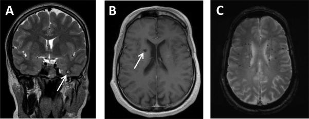
MRI appearance of vascular malformations: A: Example of classic MRI appearance of intracranial cavernous malformation (ICM) on T2+weighted imaging (Patient 3). Inferior left temporal lobe ICM showing complete low signal rim surrounding an area of mixed T2 signal. B,C: Radiographic spectrum of vasculopathies in the same patient (Patient 9): B gadolinium+enhanced T1+weighted image showing a right periventricular ICM with small area of hyperintensity surrounded by a hypointense signal, and C: T2*+weighted image showing small and scattered radiation related microbleeds shown as areas of decreased signal intensity.