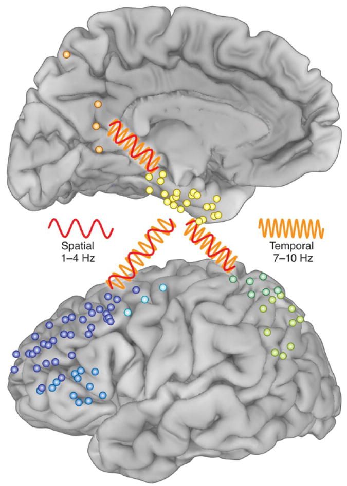
Figure 2.

Individual subdural recording sites from the patients studied in [30]; blue, prefrontal; green, parietal; orange, precuneus; yellow, parahippocampal. The red oscillation (1-4 Hz) represents coherence between brain regions during spatial memory and the orange oscillation (7- 10 Hz) represents coherence between these regions during temporal memory. Adapted from [30,31].
