
Figure 2.
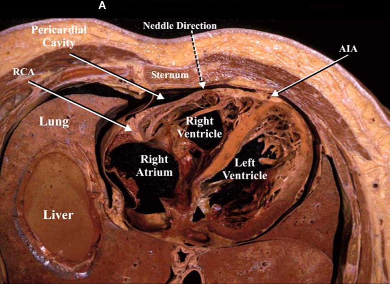
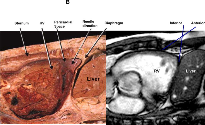
A: The figure demonstrates a cross section of the lower part of the thorax revealing the relationships of the pericardial cavity with the adjacent structures. In addition, the broken arrow is illustrating the direction of the pericardiocentesis needle. RCA, right coronary artery; AIA, anterior interventricular artery. B: midsagittal section of a cadaver revealing the pericardial cavity, the diaphragm and the contents of the abdominal cavity. In addition, the broken arrow shows the direction of the pericardiocentesis needle [fig 2 A and 2B (left panel) used with permission from Lukas et al9]