Figure 6.
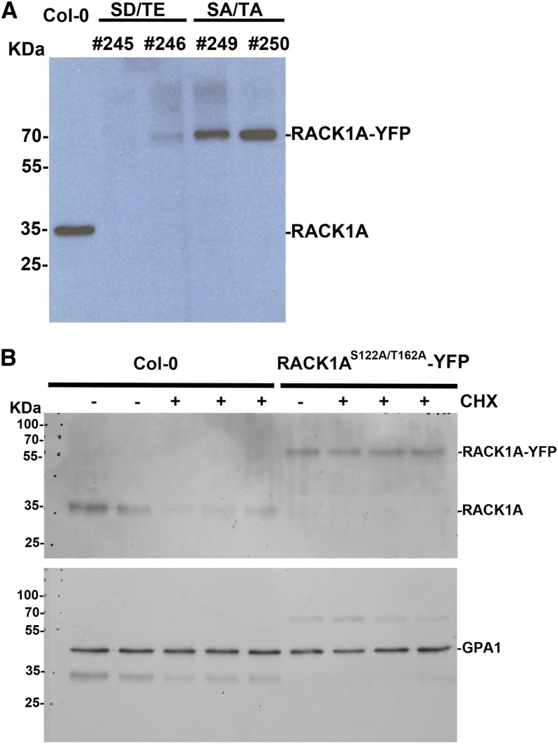
Immunoblot analysis of RACK1A protein. A, RACK1A protein in wild-type (Col-0) and transgenic plants. Total proteins were extracted from leaves of 6-week-old plants and loaded to an SDS-PAGE gel. Anti-RACK1A peptide antibodies were used for immunoblot analysis. SA/TA, RACK1AS122A/T162A; SD/TE, RACK1AS122D/T162E. B, The stability of RACK1A protein. Total proteins were extracted from 1-week-old Arabidopsis seedlings of Col-0 and PRACK1A::RACK1AS122A/T162A-YFP transgenic plants (in rack1a-2 mutant background) grown in one-half-strength Murashige and Skoog liquid medium. Anti-RACK1A peptide antibodies were used for immunoblot analysis. The same membrane was blotted with anti-Arabidopsis GTP-binding protein α subunit1 (GPA1) antibodies as a loading control. Lanes 1 and 2, Col-0 without cycloheximide (CHX) treatment; lanes 3 to 5, Col-0 treated with 70 µM CHX for 6 h; lane 6, PRACK1A::RACK1AS122A/T162A-YFP without CHX treatment; and lanes 7 to 9, PRACK1A::RACK1AS122A/T162A-YFP treated with CHX for 6 h.

