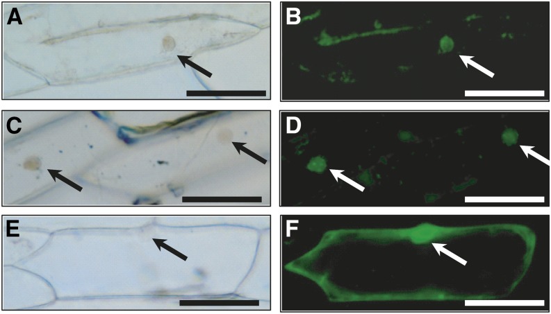
Figure 8.
Localization of NKD-GFP fusion protein nuclei of onion cells. Plasmid constructs were biolistically introduced for transient expression. A and B, NKD1-GFP. C and D, NKD2-GFP. E and F, Empty vector GFP control. A, C, and E, Differential interference contrast optics. B, D, and F, GFP fluorescence. Arrows indicate nuclei. Bars = 100 µm.

