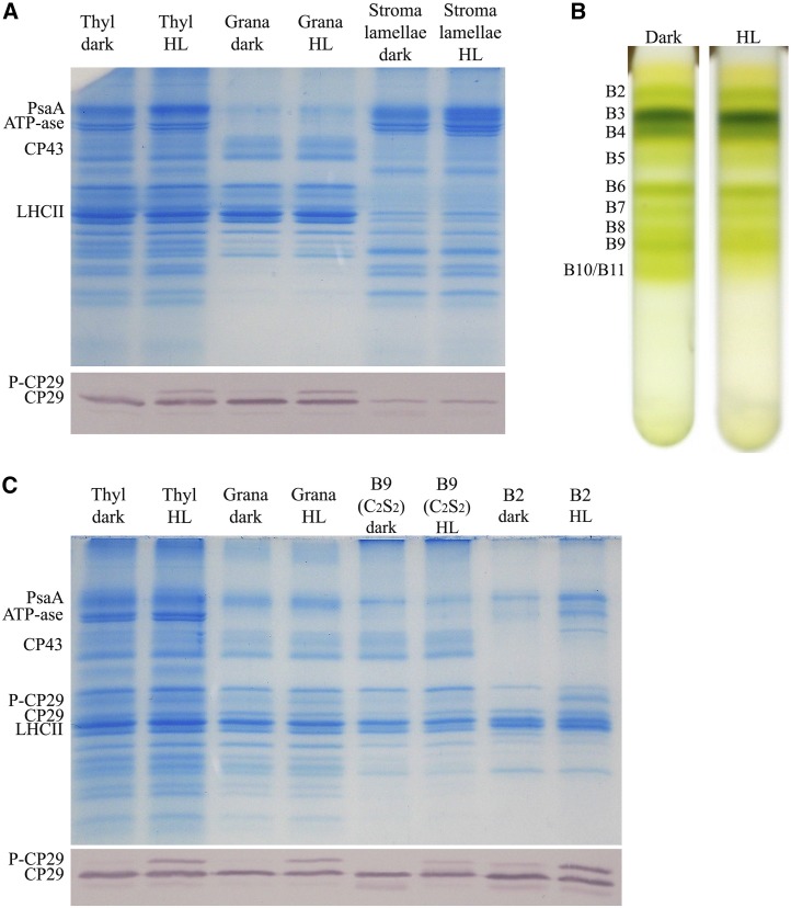
Figure 8.
Localization of P-CP29 in thylakoid membranes. A, Fractionation of thylakoid membranes for the isolation of grana and stroma lamellae membranes (see “Materials and Methods”). Thylakoids (Thyl) have been collected from dark-adapted and HL-treated rice leaves (white light, 1,000 µmol photons m–2 s–1, 30 min). These fractions have been loaded on Tris-Gly SDS-PAGE for Coomassie Blue gel staining or immunoblot analysis using anti-CP29 antibody. PsaA, PSI-A core protein of PSI. B, PSII supercomplex fractionation from grana membranes according to Caffarri et al. (2009). Thylakoids have been collected from dark-adapted and HL-treated rice leaves (white light, 1,000 µmol photons m–2 s–1, 30 min). C, Thylakoids, grana membranes, PSII-C2S2, and monomeric antenna fractions have been loaded on Tris-Gly SDS-PAGE for Coomassie Blue gel staining or immunoblot analysis using anti-CP29 antibody.

