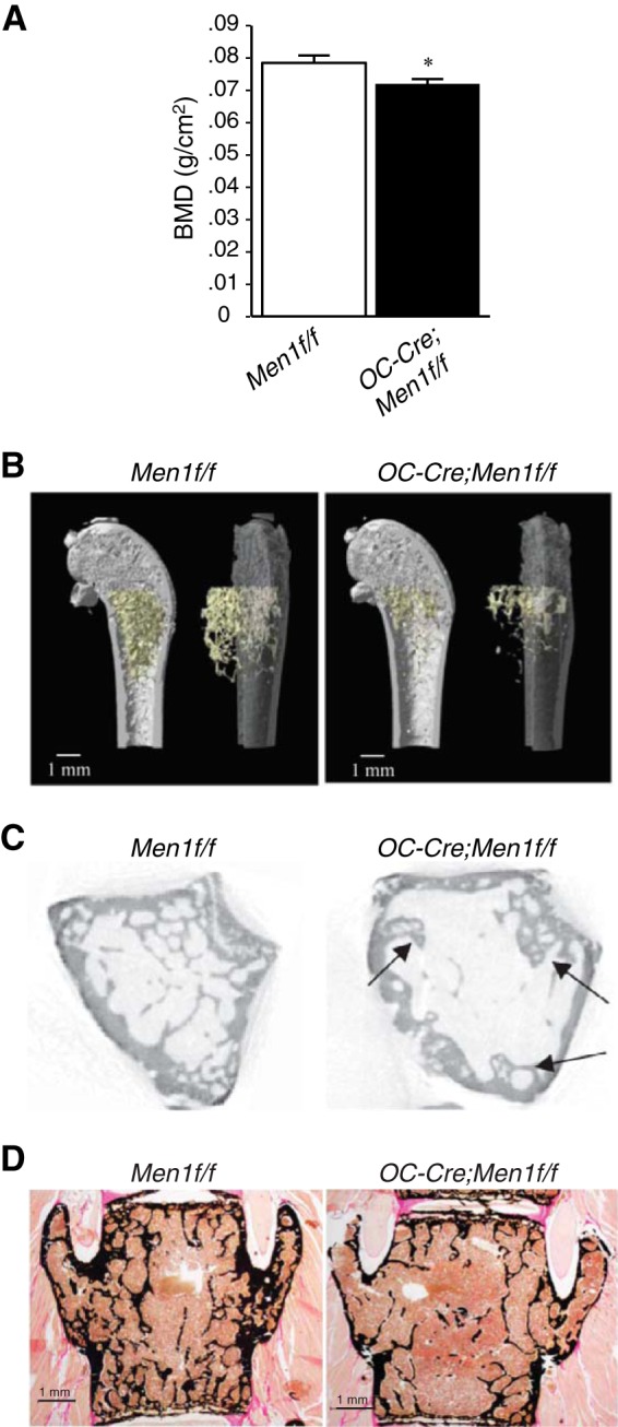
FIGURE 2.

Deletion of osteoblast Men1 leads to reduction in BMD and decreased bone volume. A, BMD (measured by dual energy x-ray absorptiometry) of femur from 9-month-old OC-Cre;Men1f/f (n = 7) and control (Men1f/f) littermate mice (n = 7). B, micro-CT analysis of distal femur of 9-month-old OC-Cre;Men1f/f and control (Men1f/f) mice (representative images are shown). C, trabecular bone was reduced in OC-Cre;Men1f/f mice compared with Men1f/f controls. Arrows indicate abnormal formation of trabeculae. See Table 1 for quantitation. Deletion of osteoblast Men1 leads to reduction in trabecular bone assessed by histomorphometry. D, representative images of von Kossa/van Gieson staining of vertebrae of 9-month-old OC-Cre;Men1f/f and Men1f/f control mice. See Table 1 for quantitation.
