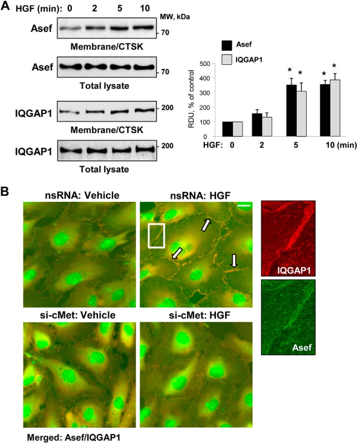
FIGURE 1.
HGF induces peripheral accumulation and co-localization of Asef and IQGAP1. A, EC were stimulated with HGF (50 ng/ml) for the time periods indicated. The content of Asef and IQGAP1 was determined by Western blot analysis of membrane/cytoskeletal fractions (CTSK) with specific antibodies and normalized to the total protein. Bar graphs depict quantitative analysis of Western blot data at membrane/cytoskeletal fractions; n = 4; *, p < 0.05 versus nonstimulated conditions. RDU, relative density units. B, HPAEC were transfected with c-Met-specific siRNA or nonspecific RNA and stimulated with HGF (50 ng/ml, 10 min). The cells were fixed and subjected to double immunofluorescence staining for Asef (green) and IQGAP1 (red). Merged images depict areas of HGF-induced protein co-localization that appear in yellow and are marked by arrows. Shown are representative results of three independent experiments; bar, 5 μm. Higher magnification insets show details of localization of Asef and IQGAP1 at the cell periphery of HGF-stimulated EC transfected with nonspecific RNA.

