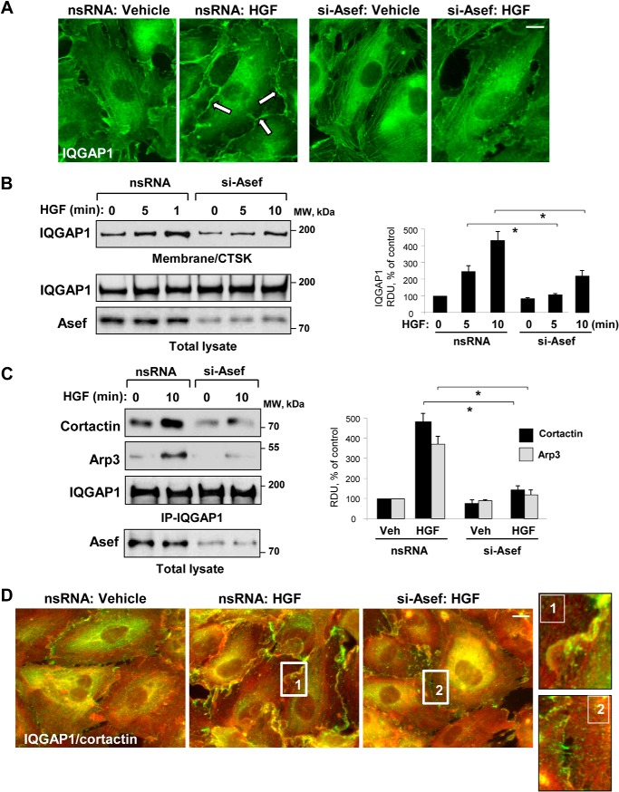
FIGURE 5.
Asef mediates HGF-induced peripheral localization of IQGAP1. HPAEC were transfected with Asef-specific siRNA or nonspecific (ns) RNA and stimulated with HGF (50 ng/ml). A, IQGAP1 localization was analyzed by immunofluorescence staining with IQGAP1 antibody; bar, 5 μm. B, IQGAP1 accumulation in membrane/cytoskeletal fraction (CTSK) was monitored by Western blot and normalized to the total protein. Bar graphs depict quantitative analysis of Western blot data; n = 4; p < 0.05 versus nonspecific RNA. C, after stimulation of control and Asef-depleted EC monolayers with HGF (50 ng/ml), co-immunoprecipitation (IP) assays were performed using IQGAP1 antibody. The presence of Arp3 and cortactin in immune complexes was determined by immunoblotting with corresponding antibody. Reprobing with IQGAP1 antibody was used as a normalization control. Shown are representative results of three independent experiments. D, HGF-induced redistribution of IQGAP1 and cortactin to lamellipodia-like structures was examined by immunofluorescence staining with IQGAP1 (red) and cortactin (green) antibody. Shown are merged images. Higher magnification insets show details of IQGAP1 and cortactin localization at the cell cortical areas of control and Asef-depleted EC upon stimulation with HGF. Shown are representative results of three independent experiments; Veh, vehicle; bar, 5 μm. RDU, relative density units.

