FIGURE 6.
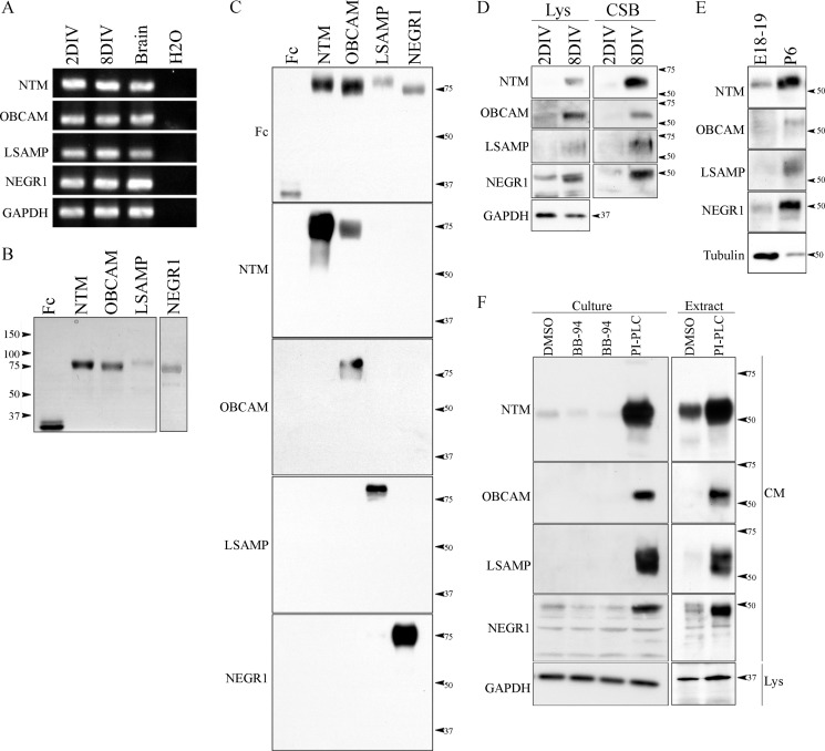
IgLON expression and processing is developmentally regulated in cortical neurons. A, reverse transcription PCR (RT-PCR) was performed in cortical neurons from two developmental stages (2 and 8 DIV). B, recombinant IgLON-Fc proteins were purified from the conditioned media of transfected HEK293T cells and analyzed by SDS-PAGE and Coomassie Brilliant Blue stain. C, specificity of commercially available IgLON antibodies was validated by Western blot against soluble recombinant IgLON-Fc proteins. D and E, developmental expression of IgLON family members was assayed in cortical neurons. Lysates (Lys) and biotinylated cell surface proteins (CSB) were collected from 2- and 8-DIV cortical neurons and lysates from the cortex of embryonic (E18–19) and postnatal (P6) stage rats. The expression of IgLON family members was determined by Western blot using commercially available IgLON antibodies. F, to validate processing of IgLON family members by metalloproteinase activity in cortical neurons, the medium of aged cortical neurons and the medium of membrane extracts from postnatal stages of brain cortex were collected. Commercially available IgLON antibodies were used to detect cleaved IgLON fragments in the medium. Anti-GAPDH and tubulin antibodies were used as loading controls. Western blots are representative of 3–4 independent experiments.

