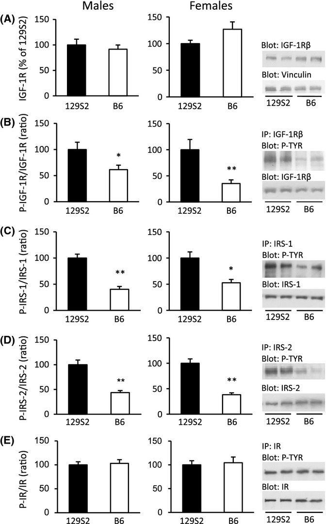
Figure 3.

Activation of IGF-1R and substrates IRS-1 and IRS-2 is higher in WT mice of 129S2 than in WT mice of B6 genetic background, in males and females. Ad libitum-fed mice (11–13 week old) were used to recapitulate pathway activation under physiological conditions. Results shown are from skeletal muscle. Representative Western blots (WB) are displayed on the right. (A) Total IGF-1R; vinculin was used as loading control. (B) Phospho-IGF-1R (P-IGF-1R), (C) P-IRS-1, (D) P-IRS-2, and (E) P-IR were detected by immunoprecipitation (IP) using specific antibodies, followed by WB using an anti-phosphotyrosine (P-TYR) antibody. Signals of activated protein were expressed relative to total protein. N = 6 per group, in males and females; *P < 0.05, **P < 0.01, Student’s t-test; error bars represent SEM.
