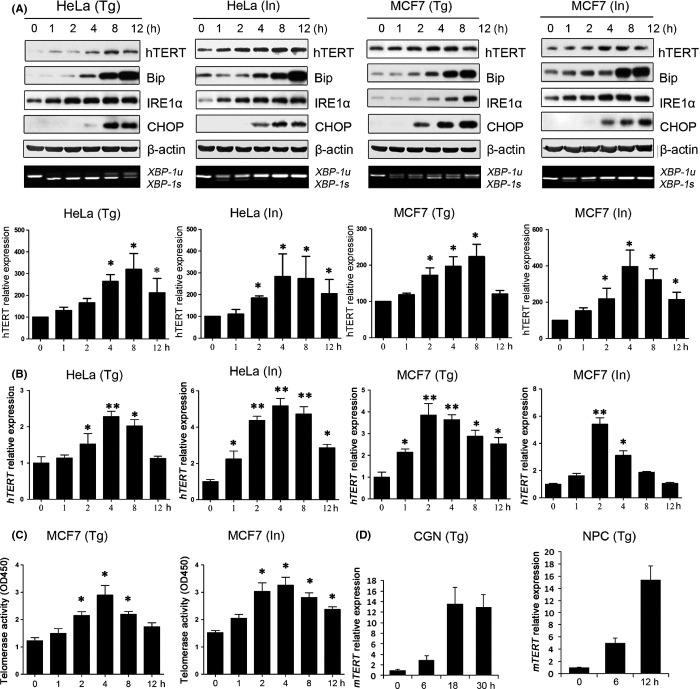
Figure 1.
Up-regulation of TERT expression under ER stress. (A) MCF7 and HeLa cells were treated with 10 μm ionomycin (In) or 2 μm thapsigargin (Tg). At the indicated time points, cells were harvested for Western blot analysis of hTERT, Bip, IRE1α, and CHOP protein expression, respectively, and for RT–PCR analysis of XBP-1u and XBP-1s mRNA expression. β-actin was used as internal control. The experiment was repeated three times and a representative result is shown in upper panel. Level of hTERT expression quantified densitometrically from three independent experiments is shown in lower panel. (B) ER stress increased hTERT mRNA expression. MCF7 and HeLa cells were treated with 10 μm In or 2 μm Tg. At the indicated time points, cells were harvested for real-time PCR analysis of hTERT mRNA expression. GAPDH was used for normalization. (C) MCF7 cells were treated with 10 μm In or 2 μm Tg. At the indicated time points, cell lysates were prepared for assays of the telomerase activity using TRAPEZE® Telomerase Detection Kit. (D) Primary mouse CGN and NPC cells were treated at indicted times. Cells were then harvested for real-time PCR analysis of mTERT mRNA expression. GAPDH was used for normalization. Data are presented as the mean ± SD from three independent experiments (*P < 0.05,**P < 0.001). Detailed experimental procedures are described in Data S1.

