Figure 1.

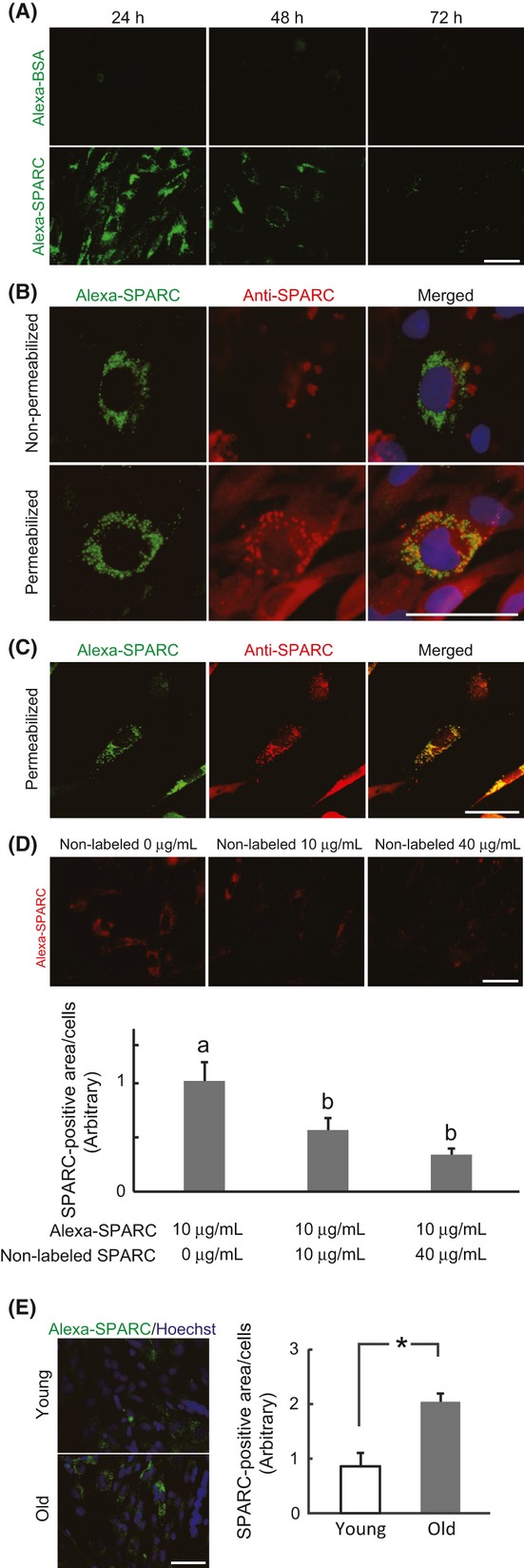
Internalization of SPARC in rat Skm-PCs and age-related changes. (A) Skm-PCs from young rats were cultured with 20 μg/mL Alexa-conjugated BSA (Alexa-BSA, above) or SPARC (Alexa-SPARC, below) for 24 h and incubated for 1 or 2 days. Times from the addition of Alexa-SPARC to Skm-PC fixation are indicated above the photographs. Scale bar = 50 μm. (B) Immunostaining of SPARC (red) was performed after incubation with Alexa-SPARC (green) with (below) or without (above) Trion-X permeabilization. Nuclei were visualized with Hoechst 33258 dye (blue). Scale bar = 50 μm (C) Skm-PCs from young rats were incubated with Alexa-SPARC (green) and immunostained with anti-SPARC antibody (red) after permeabilization; photographs were obtained by confocal microscopy. Scale bar = 40 μm. (D) Alexa-SPARC (red) was competed with non-Alexa-labeled SPARC in Skm-PCs from young rats for 12 h. Ten cells were randomly chosen, and SPARC fluorescence per cell was quantified by ImageJ. Scale bar = 50 μm. Error bars represent means ± SEM (n = 10 cells, respectively). Bars sharing the same letters are not significantly different. P < 0.05 (Tukey-Kramer’s test). (E) The photographs represent Skm-PCs from young and old rats incubated with Alexa-SPARC (green) for 12 h. Nuclei were visualized with Hoechst 33258 dye (blue). Scale bar = 50 μm. The amount of SPARC internalized into Skm-PCs was quantified by ImageJ and compared between young and old rats. Error bars represent means ± SEM (n = 3, respectively). *P < 0.05. Skm-PCs, skeletal muscle progenitor cells; SPARC, secreted protein acidic and rich in cysteine.
