Figure 1.

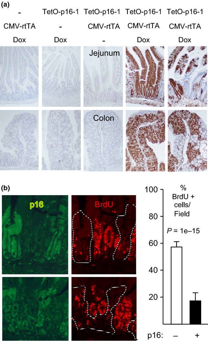
p16 induction inhibits intestinal epithelial cell proliferation. (a) Mice of different genotypes were or were not treated with Dox for 1 week, as designated. IHC for exogenous p16 (brown, 10× fields). Note strong mosaic p16 induction in the presence of both transgenes and Dox. (b) CMV-rtTA:TetO-p16-1 mice treated with Dox for 1 week and injected with BrdU. Panels (left): Two 20× fields (above, below) with co-IF for p16 (left, green; right, dashed lines) and BrdU (right, red). Graph (right): Among 1122 crypt cells scored, 19% of p16+ cells were BrdU+ vs. 58% of p16- cells. Modest BrdU signal was visible in the green filter.
