
Figure 2.
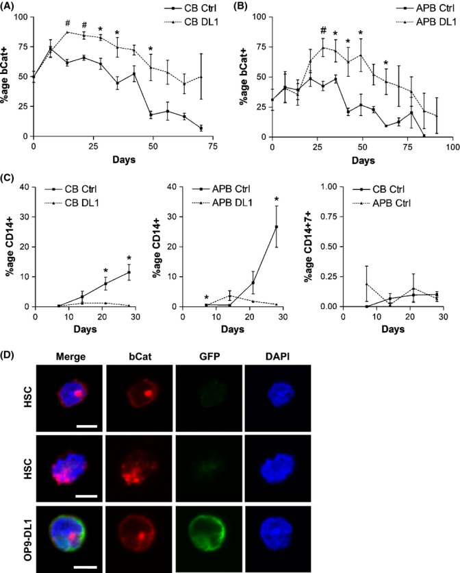
Increased expression of β-catenin in human hematopoietic stem cells (HSCs) cultured in T-cell differentiation conditions. Young (CB) and aged (APB) HSCs were co-cultured with OP9-Ctrl and OP9-DL1 stromal layers and analyzed by flow cytometry at weekly intervals. Percentage of β-catenin+ hematopoietic cells in co-cultures of (A) young HSCs (#P < 0.005, *P < 0.05; n = 3–5) and (B) aged HSCs (#P < 0.01, *P < 0.05; n = 3–7). (C) Percentage of CD14+ cells and CD14+ CD7+ cells generated in co-cultures of young and aged HSCs with OP9-Ctrl and OP9-DL1 stromal layers (*P < 0.05; n = 3). (D) Representative confocal sections depicting immunofluorescence staining of HSCs co-cultured with OP9-Ctrl and OP9-DL1 at different z-axis planes. Cells were examined for bCat (red) protein localization, and OP9 cells were identified by GFP (green) expression. Nuclei were stained with DAPI (blue). Nuclear expression of bCat was observed in young and aged HSCs during both myeloid and T-cell differentiation, and cytoplasmic bCat was also detected. Scale bar: 5 μm. CB: cord blood HSCs; APB: adult mobilized peripheral blood HSCs; bCat: β-catenin; DL1: OP9-DL1; Ctrl: OP9-Ctrl.