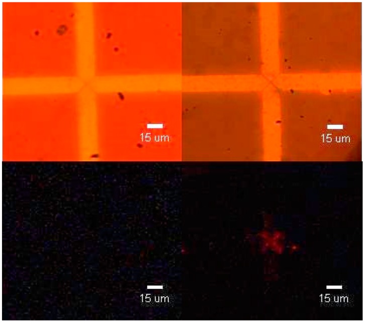
Figure 4.

Demonstration of M13 capture using triangular electrodes with a gap spacing of 4 μm. Left: Visible light (top) and UV light (bottom) images of anti-fd functionalized electrodes. Right: Visible light (top) and UV light (bottom) images of anti-fd functionalized electrodes with dielectrophoretically captured M13 phage. Conditions: Vpp = 15 V, f = 1 MHz, capture time: 30 min.
