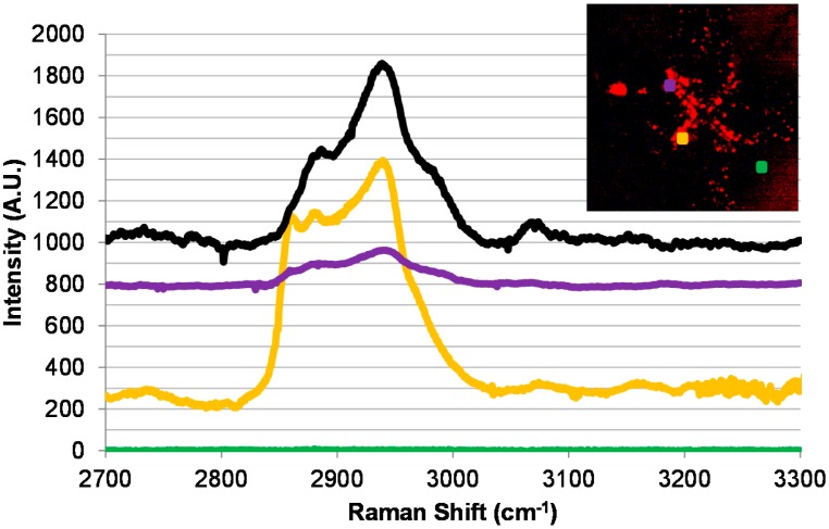
Figure 5.
Micro-Raman spectra collected from an anti-fd functionalized microelectrode with captured M13 phages bound to biotinylated-anti-fd reacted with ExtrAvidin-Cy3 prior to photobleaching. The inset in the top right is a magnified image of the collection presented in Figure 4. Black line (positive shifted by 1000 A.U.): a photobleached sample of ExtrAvidin-Cy3 on Silicon. Purple line (positive shifted by 800 A.U.): a scan from the top left point in the inset (indicated by the purple dot). Yellow line (positive shifted by 250 A.U.): a scan from the bottom left point in the inset (indicated by the yellow dot). Green line: a scan from the bottom right point in the inset (indicated by the green dot).

