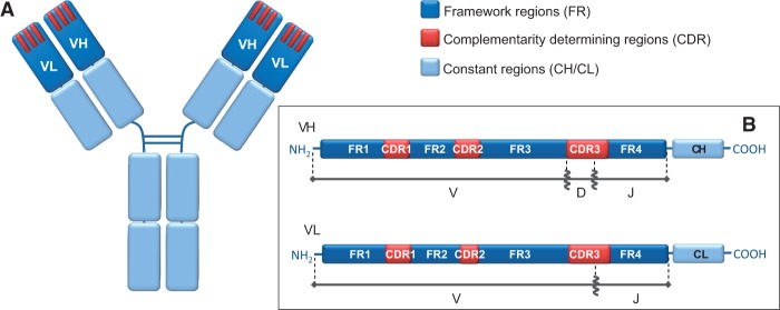
Fig. 1.

Antibody structure: (A) General schematic representation highlighting the apical position of the variable heavy chain (VH) and variable light chain (VL) domains. CDRs are represented as three red bars for each variable region. (B) Linearized representation of the variable regions, with their respective original germline gene fragments.
