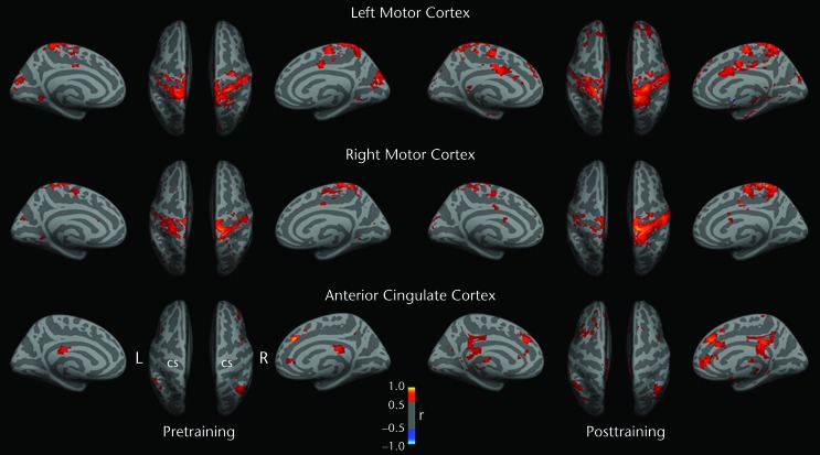
Figure 3.
Functional connectivity (FC) results projected onto inflated medial and superior cortical surfaces in standard Montreal Neurological Institute (MNI) template space. At baseline, greater FC strength was observed when the left (L) motor cortex leg representation was seeded than when the right (R) motor cortex leg representation was seeded. After training, increased FC was observed for the leg areas of both primary motor cortexes and multiple cortical regions. When the right motor cortex was seeded, connectivity strength appeared to show increased lateralization after training; this feature was not apparent for the left motor cortex. Warmer colors represent higher positive correlations between seed activity and voxelwise brain resting activity. The right anterior cingulate cortex (MNI coordinates: x=5, y=34, z=28) was used as a control site for changes in nonmotor cortical regions. This region was shown by resting-state fMRI to be functionally connected to nonmotor prefrontal cortical regions related to attentional processing and executive function.32 The spatial pattern of connectivity was largely unchanged after training. CS=central sulcus.

