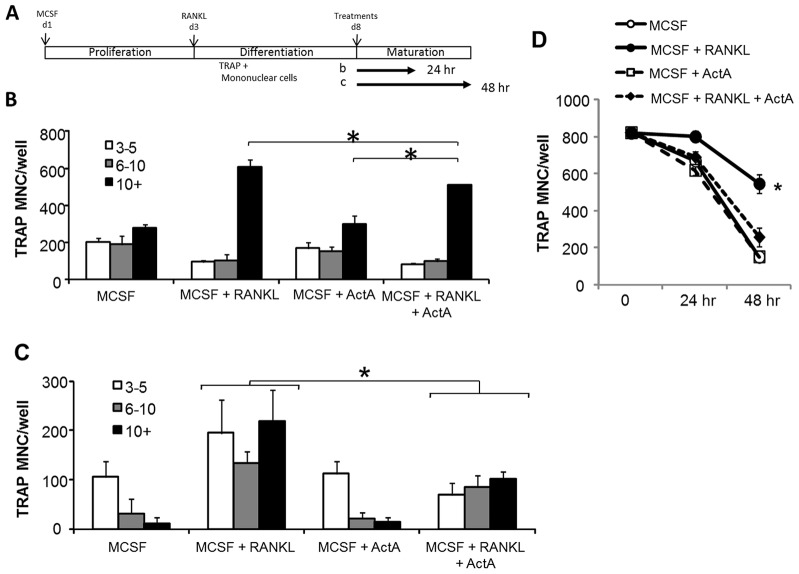
Fig. 4.
ActA suppresses mature osteoclasts. (A) Schematic of expansion, differentiation and maturation of murine OCLs. Murine bone marrow cells were expanded for 3 days in the presence of 100 ng/ml MCSF, followed by trypsinization and plating in the presence of 20 ng/ml MCSF plus 50 ng/ml RANKL for another 5 days. On day 8, medium containing MCSF plus RANKL was aspirated and the cells treated with either MCSF alone or in combination with RANKL and 50 ng/ml ActA for 24 hours (B) or 48 hours (C). After 24 hours (B) or 48 hours (C) treatment, medium was aspirated and cells were stained for TRAP. TRAP+ MNCs and the number of nuclei per cell were counted (3–5 nuclei, white bars, 6–10 nuclei, gray bars; greater than 10 nuclei, black bars). Bars represent the mean±s.d. number of cells from triplicate wells in multiple experiments. (D) TRAP+ MNCs from day 8 MCSF plus RANKL cultures were harvested (t = 0), and treated with MCSF alone, or in the presence of ActA, RANKL or both for 24 or 48 hours. Cells were TRAP-stained, and the total number of TRAP+ MNCs containing at least three nuclei was counted in triplicate wells. *P<0.05 compared with MCSF alone. Analysis is representative of experiments performed in triplicate.

