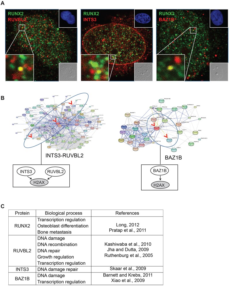
Fig. 5.
RUNX2 association with RUVBL2, INTS3 and BAZ1B in vivo. (A) Immunofluorescent staining of RUNX2 (Alexa Fluor 488, green) with RUVBL2, INTS3 or BAZ1B (Alexa Fluor 555, red) in Saos2 cells was analyzed by confocal microscopy (z-sections were 0.2 µm thickness; images were acquired using a 63x objective with oil immersion; 1.4 numerical aperture). Insets show differential interference contrast (DIC, lower right) and DAPI images (upper right). (B) The functional protein networks of RUVBL2, INTS3 and BAZ1B were analyzed using STRING (version 9.0). Boxes below the network diagrams show a simplified version of the model. (C) Reported biological functions of RUNX2, RUVBL2, INTS3 and BAZ1B, with references.

