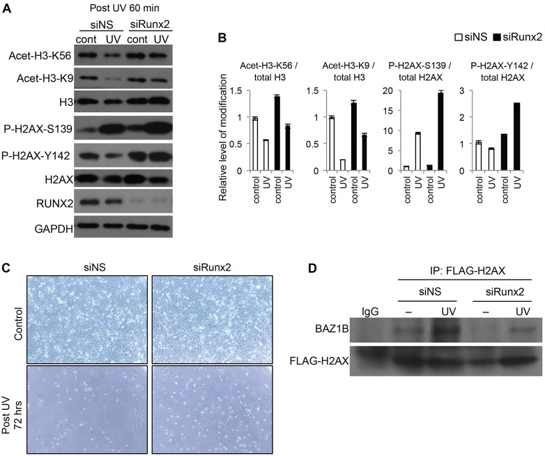
Fig. 8.
RUNX2-dependent histone modification in response to UV. (A) Saos2 cells transfected with nontargeting siRNA (siNS) or RUNX2-targeting siRNA (siRUNX2) were non- irradiated (cont) or were UV irradiated (300 J/m2). Then, cells were further incubated for 60 min, and K56 and K9 acetylation of histone H3 and S139 and Y142 phosphorylation of histone H2AX were analyzed by western blotting. (B) The graph shows quantification of the western blot data shown in A performed using Image J software. Each band from acetylation or phosphorylation of H3 or H2AX was measured and normalized to total H3 or H2AX levels. The image represents the analysis of at least three independent western blots. Data show the mean±s.d. (C) Saos2 cells transfected with siNS or siRUNX2 were non-irradiated (Control) or UV irradiated (UV, 300 J/m2), and images of cells were analyzed by Nikon phase-contrast microscopy (10×). (D) Immunoprecipitation (IP) analysis of UV irradiated (UV, 60 min) or non-irradiated (–) Saos2 cells co-transfected with FLAG–H2AX expression plasmid and the indicated siRNA using anti-FLAG antibody. Immunoprecipitation with normal IgG was used as a control.

